


Keratoconus and Its Treatment Using Lenses and Surgery
The eyes, a network of two million moving parts, is inarguably the most important sense organ of the human body. Whether gazing into the eye of a loved one, steering down a car through a mazy road, or going through a gallery of colorful artworks, the power, and importance of the eye cannot be underestimated.
It is your window to the world, allowing you to experience and savor every moment of your existence. In fact, according to Zeiss, we perceive 80 percent of our impressions using what we see.
The eyes are truly amazing organs and can capture and interpret an average of one-million pulse signals per millisecond, transmitting to the brain in the fraction of a second. However, that important organ of the body is threatened daily by the things we do, eat, come in contact with, or even some undetermined factor like our genetic makeup. The eyes, just like other sense organs present in the human body, are subject to injury and stress and can lose its strength and full potential if not properly taken care of. To contain some of these posed threats, nature has put in place some design such as the reflex batting of the eyelids when an object is coming in its direction, or the provision of the eyelids and eyelashes to keep out dust.
That said, a number of eye defects can’t be placed in check by these natural provisions. One of such is keratoconus.
Keratoconus, simply put as a cornea in a coned shape, is a progressive eye defect affecting the cornea and front window of the eye, which results in poor vision that often cannot be fully corrected with glasses.
The Science Behind Keratoconus
The direct cause of keratoconus isn’t known. However, the defect is birthed by the weakening of tiny fibers of protein in the eye called collagen. The collagen fibers help hold the cornea in place, preventing it from bulging. When these fibers become significantly weak, they can no longer hold the cornea in place, and as a result, leave the cornea to become cone-shaped progressively.
 A decrease in the level of protective antioxidants in the cornea can also cause keratoconus. Like the exhaust of a car or that from a factory, the cornea cells are designed to produce damaging by-products. In general, antioxidants are meant to get rid of them and consequently protect the collagen. If this is so, low levels of antioxidants can leave collagen fibers unprotected, leaving the cornea to bulge out.
Causes of Keratoconus
This defect is suspected of running in families, through generations after generations. Consequently, if you have it, it is recommended that you have your children checked starting at age 10. For some people with certain aiding medical problems such as Downs Syndrome and allergic conditions, the defect progresses rapidly.
In some quarters, incessant rubbing of the eye is also suspected to be a culprit. Save from that, chronic irritation, overexposure to ultraviolet rays, and history of poorly fitted contact lenses also aid the development of keratoconus.
In most cases, the defect starts in teenage years. Although it starts in childhood, people up to age 30, and people aged 40 and over can develop keratoconus. However, these are rather exceptions than the norm. The shape of the corner can change very quickly or over a long stretch of years.
Symptoms of Keratoconus
The symptoms of keratoconus aren’t in all cases clear-cut, and could sometimes need a diagnosis to confirm its presence. Be that as it may, the general symptoms signaling the presence of the Keratoconus defect include:
- Lights streaking / Glare
- In advanced cases, seeing double or triple ghost images.
- Double vision when looking at an object with only one eye.
- Objects placed both far and near appear distorted.
- Blurry vision while driving. Especially while doing so at night.
- Bright lights appear like they have halos around them.
- A sudden change of vision in one eye
Diagnosis of Keratoconus
Even with the aforementioned symptoms, it is highly recommended that some tests are carried out to confirm the presence of this defect. These tests are to be performed by a keratoconus eye specialist (ophthalmologist or optometrist).
The specialist is expected to review your ‘family’s medical history and might take some extra tests to determine several other details like the shape of your cornea. The tests taken to diagnose keratoconus include:
Keratometry: In this test, the doctor focuses a circle of light on the cornea and measures the reflection of the light in a bid to determine the basic shape of the cornea. The corneal curvature helps in determining the power of the cornea. This measurement can be determined manually or via a number of automated methods.

Eye Refraction: In this test, special equipment is used to check the eyes for vision-related problems by taking measurements of the eye. The doctor may require that you look through a device that comprises of wheels of different lenses, also known as a phoropter. Consequently, this helps to arrive at the conclusion of which arrangement gives you the sharpest vision. In some cases, the eye specialist may make use of a retinoscope to evaluate your eyes.
Computerized Corneal Mapping: This employs the use of special photographic tests such as corneal topography and optical coherence tomography to record images of the cornea. This, in turn, creates a detailed map of the ‘cornea’s surface. The tests do not stop at this. They also assist in measuring the thickness of the cornea.
Wavefront Aberrometry (HOA) Mapping: With Aberrometry testing we can measure the advanced visual errors known as higher-order aberrations (HOAs). The higher-order aberrations (HOAs) commonly seen with-in keratoconus cause distorted vision – difficulty seeing at night, glare, halos, blurring, starburst patterns or double vision (diplopia).
Slit-lamp Examination: This eye examination is done to evaluate the shape of your cornea as well as other potential eye problems that might have erupted. During this examination, the doctor directs a vertical beam of light on the surface of the eye by the use of a low-powered microscope.
Treatment of Keratoconus
In general, treatment starts with the use of eyeglasses. If these glasses do not provide adequate correction, then special lenses such as the rigid glass permeable contact lenses are recommended. In advanced cases of the defect, and only when medically necessary, surgical procedures are best suited.
The use of Keratoconus Lenses:
The irregular shape of the cornea worsens image quality and consequently necessitates the need for vision correction in most patients. The type of lens to be used should be custom-tailored to each patient. As a rule of thumb, the use of lenses would suffice for patients in the early stages of keratoconus.
However, as the defect worsens, patients have to ramp up regularly to use stronger and stronger lenses. For the most part, contact lenses function by creating a smooth and artificial surface on the front of the eye that improves its ability to bend the light entering it. Frequent changes in the shape of the cornea may necessitate the need for lenses with a different shape and power. Special contact lenses have been developed specifically for those suffering from keratoconus. Some of these include:
Soft Contact Lenses: They are not a typical option for Keratoconus patients. However, some soft contact lenses provide vision improvement for those who cannot cope with hard lenses. They provide significantly less visual clarity. Hence, the need for patients to opt for a compromise that stands somewhere comfortably in between optimum vision and comfort.
Rigid Gas Permeable Lenses: They are the most commonly prescribed contact lens type for correcting keratoconus. They are excellent for the ‘eye’s health, as they allow the cornea to breathe oxygen through its material. They can be customized and designed solely for the unique shape of the affected cornea. They are also easy to apply, remove, and care for. They provide great vision correction. However, some patients cannot tolerate wearing them for long periods of time.
Piggyback lenses: It is a technique where soft contact lens and a corneal lens (or hybrid lens), is used in tandem. In this method, the soft contact lens is placed directly on the cornea, and a corneal GP lens is made to sit on top of the soft lens. Although employing this method has been found to be more tasking, in most cases, it saves the patient from problems of irritation.
Hybrid lenses: Hybrid lens is a specialized contact lens that blends the advantages of a rigid gas permeable lens and that of a soft contact lens. This lens type gives its users the visual acuity of a rigid gas permeable lens and the comfort of a soft contact lens. It is a viable option for you if you are not comfortable wearing a rigid gas-permeable contact lens.
Scleral lenses: They are large-diameter GP lens, about the size of a nickel to a quarter. It is designed to perfectly vault over the cornea and rest on the sclera – which is the white part of the eye. Due to its size, the lens bowl is filled with non-preserved saline before it is placed on the eye. Initially, scleral lenses wearers may find applying and removing it challenging, but with proper training, the insertion and removal become second nature. The exceptional vision and comfort it provides far outweigh any of the insertion challenges.
Custom Scleral Shells: Similiar to standard scleral lenses, they are also large-diameter lenses, often larger, but are designed using digital Optical coherence tomography (OCT) and/or EyePrintPRO™ custom prosthetic scleral lens moldings. With the precise OCT data and custom molding, the scleral shells can be designed to provide exact contour match. With Precision Vision Rehabilitation Prosthetic Replacement of the Ocular Surface Ecosystem (PVR PROSE), EyePrintPRO™, and other custom scleral shells great vision and comfort can be obtained.
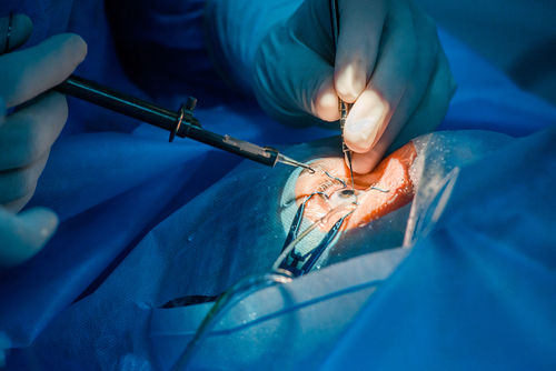
The Surgical Approach to Keratoconus:
There are several other new methods used to tackle keratoconus. The most common is a corneal transplant but is often not truly needed. However, the surgical approach of a corneal transplant is essential for severe cases when there is a concern of the cornea rupturing. To truly halt the progression of keratoconus Corneal Cross-Linking (CXL) is needed. With the use of eyedrop-medication and ultraviolet (UV) light from a special machine Corneal Cross-Linking (CXL) makes the tissues in the cornea stronger. The goal is to keep the cornea from bulging and halt the progression of keratoconus.
Corneal Cross-Linking (CXL): Approved by the FDA in 2016 Corneal Crosslinking (CXL) uses a riboflavin ophthalmic solution (Photrexa, Avedro) to strengthen the cornea and halt the progression of keratoconus. Studies have shown that Corneal Crosslinking (CXL) has been highly successful in halting keratoconus, and is a safe and effective treatment for keratoconus. While it does stop keratoconus, it does not necessarily provide improvement to vision. Most often CXL is combined with a multitude of vision correction options: scleral lenses, intrastromal ring implants (Intacs), PRK, phakic IOL implantation, or conductive keratoplasty (CK).
With the introduction Corneal Cross-Linking (CXL), early childhood detection of keratoconus is crucial. When diagnosed with keratoconus at an early stage Corneal Cross-Linking (CXL) can stop the progression and prevent permanent vision impairment.
Corneal Transplants: A transplant is the last resort, applicable usually in cases where all methods have been exhausted and vision improvement and comfort cannot be achieved for the patient. Corneal tissues are harvested from deceased organ donors just within hours of their death. Thereafter, they are tested to ensure they are disease-free and suitable for surgical procedures. Although transplants can last decades with adequate care, results from person to person may vary.
The two most commonly employed methods are the Penetrating Keratoplasty and Deep Anterior Lamellar Keratoplasty methods.
Penetrating Keratoplasty: This is the traditional practice of surgical remedy used for KC. In this procedure, a trephine is used to harvest the full thickness of the cornea, that is, from epithelium to endothelium. The ‘patient’s damaged cornea is then replaced with similarly sized tissue harvested from the donor. A series of individual sutures or single continuous suture is done to hold the transplant in place. As time goes by, eyes heal, and vision improves, the stitches are then removed. The operated patients are then usually given eye drops for an extended period to ensure that the transplanted tissue is not rejected.
 In most recent cases, the removal of the damaged cornea, and preparation of the harvested cornea from the donor are done via the use of femtosecond laser as opposed to using a trephine. The laser is a precise-cutting instrument that can ensure better-customized patterns in both the recipient of the harvested cornea and its donor. If employed, faster wound healing and stronger bonds are promised, ultimately resulting in lower astigmatism risks.
Deep Anterior Lamellar Keratoplasty (DALK): DALK, a less invasive surgery procedure, has become a popular alternative among expert eye surgeons. In this method, the outer layers of the affected cornea are removed and replaced, but Descemet’s membrane and endothelium are left untouched. It ensures quicker recovery time and a lesser probability for graft rejection.
It goes without mentioning that whatever the surgical method employed, it is still likely that you would need some vision correction with the use of contact lenses. In some cases, the graft might be rejected, and the surgeon would be needed to perform a repeat surgery. Infections are also a potential complication.
Conclusion
As mentioned earlier, the eye is a very important organ of the human body and quite delicate, to say the least. As a result, individuals should take good care of their eyes and go for a check or diagnosis if they notice any abnormalities. Many patients have their heart skip a beat when they are diagnosed with keratoconus. They get even more scared when the thought of a corneal transplant crosses their minds. However, there would not be a need for a transplant or surgical procedure in most cases, as this is only a last resort solution.
With that being said, it is imperative that patients know that with the introduction of newer scleral lenses and the advent of Corneal Cross-Linking (CXL), corneal surgical procedures are often not needed.