


Cornea Transplants and Scleral Lenses for Keratoconus
Advantages and Disadvantages of Cornea Transplant and Scleral Lenses For the “Treatment” of Advanced Keratoconus
Picking the right treatment for any disease plays a vital role in recovery. There are several different treatment methods for advanced keratoconus. But in this article, we will try to help you understand just two treatment types better – cornea transplant and scleral lenses.
These two keratoconus treatment options are among the most popular treatment options. They are regularly touted as the easiest and most effective treatment methods in some quarters. But when you are done reading this article, you’d be well informed to form your own opinions about them.
Keratoconus is a progressive medical condition of the eye that affects the cornea. The cornea thins and bugles out like a cone, leading to vision problems. Keratoconus treatment helps to halt the progression of the disease. And in some cases, they help keratoconus patients with partial visual rehabilitation. New treatment methods are still being discovered, so there’s no particular best treatment or most effective. It all depends on you.

For non-medical students or practitioners, the definition and explanation of keratoconus given above is still very likely to leave you confused. Let me break it down for you to understand much better.
Keratoconus is a disease which makes the cornea of the eye, which is the transparent tissue in front of the eye, to bulge outward. When a person suffers from keratoconus, the clear dome-shaped tissue that covers the eye, particularly the cornea, thins, and bulges outward into a cone shape.
The cause of this disease is yet to be known, and it affects a small population of the world. The occurrence of keratoconus can be found in about one in every two thousand people*.
Causes of Keratoconus
There is no finite cause of Keratoconus known. However, research suggests that it is caused by an enzyme imbalance within the cornea, which weakens the cornea. This imbalance exposes the cornea to the risk of oxidative damage by free radical compounds.
 Keratoconus is also linked with excessive rubbing of the eyes, overexposure to the sun’s UV rays and chronic eye irritation.
Signs and Symptoms of Keratoconus
The cornea is the most critical lens of the eye. It is responsible for the bending of light, which is the primary determining factor for sight. So, when it is affected, the sight itself is affected. For the cornea to properly transmit and focus light, it needs to remain clear and be of perfect shape. All of which are affected when suffering from keratoconus.
The cornea of both eyes is usually affected when suffering from keratoconus, although the signs may be more noticeable in one than in the other. Symptoms typically become noticeable in adolescence, or the early stages of adulthood. It is very common for it to grow progressively worse 10-20 years before eventually slowing down.
Vision changes can vary from one person to another, and its severity can range from mild vision loss to more severe vision loss. Severe vision loss that makes it next to impossible to see, even with aid from standard corrective lenses.
Keratoconus patients start suffering from high sensitivity to light and glares, and their vision also begins to get blurry.
Treatment Options for Advanced Keratoconus
There are several treatments used for Keratoconus, but when medically necessary, Keratoconus can be treated by Penetrating Keratoplasty (PK), and its cousin – Deep anterior lamellar Keratoconus (DALK). In most cases, Scleral lenses are very helpful to provide better vision, but not to stop the progression. Corneal Cross-Linking (CXL) treatment is invasive procedure to stop the progression of keratoconus, but does not necessarily improve one’s vision. Many patients will still need some type of vision correction after Corneal Cross-Linking (CXL).
For the scope of this article, we will be focusing on just two Keratoconus treatment options – corneal transplants and scleral lenses.
Corneal transplant:
A corneal transplant is not a simple procedure, and should truly only be done when medically necessary – not just to improve vision. Patients can face significant risks with a corneal transplants – corneal rejection and infections. Additionally, post-transplant medications can have side effects that can cause glaucoma/high eye pressure.
Corneal transplant serves as the last resolve to be considered, and it is also called penetrating keratoplasty (PK or PKP). Almost all keratoconus patients that have a corneal transplant will still require contact (scleral) lenses or glasses will still be needed for clear vision.
For those suffering from advanced keratoconus and at a high risk of corneal perforations or corneal rupture, Deep Anterior lamellar Keratoconus (DALK) and Penetrating Keratoplasty are seen as some of the best treatment options. Once visual acuity becomes unacceptable a scleral lenses is the next step.
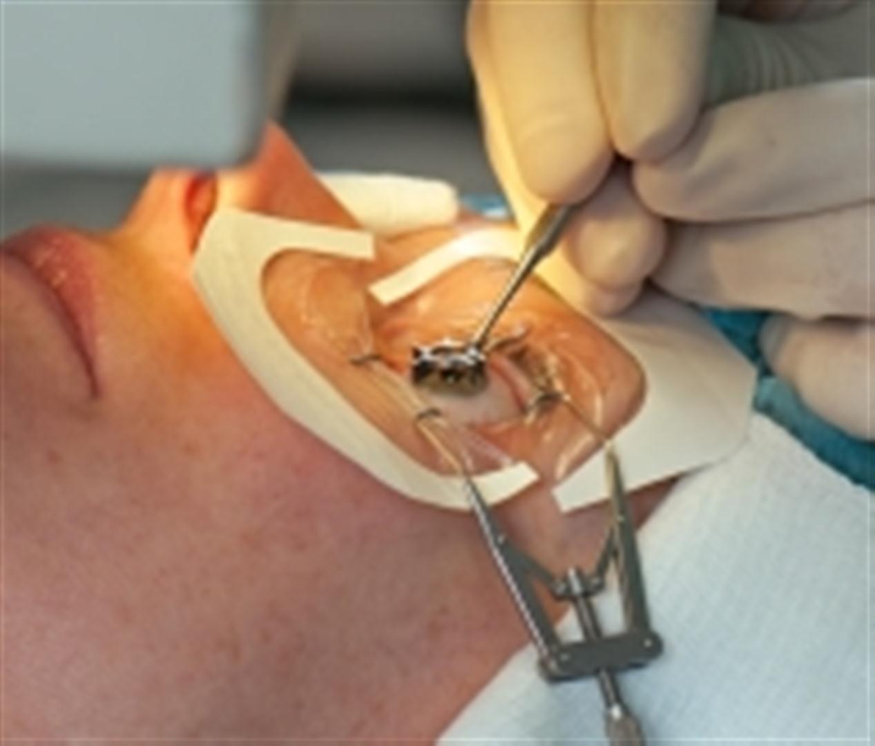
In this procedure, the doctor removes a full thickness portion of the central cornea and replaces it with the donor tissue. The DALK is then inserted to preserve the inside lining of the cornea (endothelium).
The results of these surgical operations are often reported to be relatively good when compared with the disappointing results from transplantation for other issues. A corneal transplant can, however, have possible complications which may include poor vision, graft rejection, astigmatism, infection, and the inability to wear standard contact lenses.
As a matter of fact, many patients suffering from advanced keratoconus tend to be inappropriate candidates for both DALK and PK procedures. Nearly all patients are young. Some as young as fifteen, which results in technical challenges during the surgery, and as a result of this, post-surgical care becomes even more difficult.
 Another challenge faced is that young eyes tend to be phakic: many of them start developing cataracts a few short years after the transplant. Due to this, their lens may have to be extracted, posing a big risk to the health of the graft.
Children already suffer poorer graft than adults, so they suffer greater risks. And even if the grafts were statically identical, it is still very likely that young patients will outlive their operation, and they would need it to be redone. Due to the poorer results gotten when operations are carried out for a second or third time, several patients who were thought to be doing great after both procedures will likely run into problems shortly.
This is specifically true because patients with a serious disorder of the ocular surface tend to suffer advanced keratoconus and many of these conditions are made worse by DALK/PK, their big incisions, sutures and the neurotrophic corneas that they birth. Because the stroma at the junction between the recipient and the graft never properly heals, the wound healing problems will continue beneath the surface of the ocular.
This process poses serious difficulties, and they are fundamental problems related to PK and DALK, so changes to the surgical technique or instrumentation ‘wouldn’t make it any better. There are better surgical alternatives. Ones that don’t require that the patient’s cornea be exchanged or replaced with that of the cornea.
Cornea transplant on a broad scale is the best treatment for advanced keratoconus when there is concern of rupture. Due to the risks of treating keratoconus by way of corneal transplant, there’s a strong push that keratoconus should first be treated in its mild stages with Corneal Cross-Linking (CXL) and scleral lenses, before a transplant is required.
Scleral lenses:
Keratoconus patients at any age or stage of the medical condition can seek relief using treatment options, to correct vision other than a corneal transplant and related surgical procedures. At the very least, with the use of contact lenses, patients can enjoy vision improvements, and keratoplasty can be avoided.
Scleral contact lens technology has brought significant improvements to the visual enhancements that patients of keratoconus can obtain. So much that even patients living with severe cases of keratoconus can see better – achieving as high as 20/20 vision, no glare, no halos, no ghosting, no double vision, etc.
Scleral lenses are actually best suited for use by patients with steep corneas because it has a large diameter lens that vaults over the cornea. This vaulting provides a fluid layer between the lens and cornea, which reduces eye irritation that is commonly experienced with lens-to-cone contact.
When patients are diagnosed with keratoconus by a physician, they should first be advised to go with mild treatments such as using the scleral lens. Except in cases where there is concern of rupture and other medical worries. Even with severe central scarring, patients have a good possibility to enjoy improved vision with the use of a scleral lens.
Basically, instead of patients automatically opting for corneal transplant as a first treatment option, it should be determined whether they can achieve good vision using a scleral lens.
Whether the condition is mild, moderate, or severe, scleral lenses can be fit on any patient with keratoconus. As a matter of fact, many patients may eventually end up using scleral lenses after a corneal transplant, so first determining whether scleral lenses will provide an improved vision could help a good number of patients to avoid a transplant altogether.
Scleral lenses are also very helpful in cases of advanced keratoconus, where there are irregular changes to the shape of the cornea. Unlike traditional lenses that rest on the cornea, scleral lenses sits on the white part of the eye (the sclera), and vault over the cornea without coming into direct contact with it.
The space left for the rigid structure of the cornea allows the user more comfort, and it still maintains its efficiency. After fitting a scleral lens, patients will need to follow up with regular check-ups to ensure that the lens fitting remains satisfactory. It is important that the lens is fitted by a professional because an ill-fitted lens could damage the cornea.
In a study in Antwerp to show the success and failure rate of the use of scleral lenses to improve the eyesight of patients with advanced keratoconus, 75 patients were tested.
Out of this study group, 8 of the patients underwent transplant surgery, while 12 patients had no scleral lens fitted in, while three patients were fitted with hybrid or corneal lenses. Fifty-one out of the total number of 75 patients were fitted with scleral lenses.
The mean gain in visual acuity (lens vs. spectacle-corrected visual acuity) was 0.54 ± 0.18 (decimal fraction, Snellen eye chart). Seven eyes were lost to follow-up, four eyes abandoned wearing the scleral lens because of an inability to handle the lenses, and 40 eyes wore the lenses at their last follow-up visit, with a mean follow-up interval of 30.15 ± 12.83 months.
The result of the study showed that 40 of the 51 patients suffering from severe keratoconus were successfully treated (they had better vision) when wearing scleral lenses for an extended period. These were patients who would have been forced to undergo corneal transplant due to their severe case of keratoconus condition.
This shows just how effective scleral lenses can be in treating keratoconus before other options like surgery should be considered.
Conclusion
Scleral lenses provide a pleasant and safer alternative to corneal transplant. But for very severe cases of keratoconus, where there is next to total blindness, a corneal transplant is advised. This is because it is one of the only ways to bring back sight.
As long as corneal transplant isn’t absolutely required, scleral lenses are advised, but having read this article, you should feel comfortable enough to choose whichever method is best suited for you. However, we’d strongly advise that you make that choice with the guidance of a professional eye doctor.